Blogging Part III (3.2)

In such organism as myself, the mouse, I contain a endocrine system which is referring to the collection of cells, glands, and tissues of an organism that secrete hormones directly into the bloodstream to controls my physiological and behavioral activities. With respect to me as a cardiac muscle I interact with my receptors to such other endocrine cells due to my receptors which can result in increase or decrease in my heart size. As a result we can conclude to this statement that there should be a regular balance between how much endocrine hormone cells I interact with.

In the fetal and embryonic stages of development, testosterone promotes the development of the penis and scrotum and the formation of the structures involved in sperm production. Hence when I interact with testosterone I bound to the albumin and globulin proteins as well as those that do not bind cause physiological changes.

Blogging Part III (3.1)

Muscle cell vs. Nerve cell

The contraction of smooth muscle is controlled by the brain through the Autonomic Nervous System (ANS). The ANS is the part of the brain which controls the movement of involuntary muscles such as smooth muscles, widening and narrowing of blood vessels and beating of the heart.

Image

Smooth muscle cells, is the most common type of cells found in the walls of blood vessels. In this region, they are termed vascular smooth muscles that either contract or relax to control the diameter of the vessel. These cells are small with thin and thick filaments. Smooth muscle cells are divided into two sub groups- single unit (or unitary) and multiunit smooth muscles. Some of these are able to act independently of each other while others need to communicate with each other via gap junctions. A gap junction or nexus is a specialized intercellular connection between multitudes of animal cell-types. It directly connects the cytoplasm of two cells, which allows various molecules and ions to pass freely between cells. Gap junctions composed of tubes made up of channel-forming proteins called connexins that together form connexons which permit the passage of inorganic ions from cell to cell. These gap junctions allow for electrical communication between neighboring cells and open and close according to changes in Ca2+ levels and pH. Calcium ions enter the cell, binding to an enzyme complex on myosin. This complex transfers a phosphate group from ATP to myosin which activates it. Myosin then forms cross bridges with actin resulting in muscle contract. Removal of calcium results in dephosphorylation of myosin that makes it inactive and relaxes the muscle.

Image

Smooth muscle cells need to be able to facilitate communication in order to synchronize contracts in blood vessel walls that then allow for control of the blood volume and pressure flowing through it.

Nerve cells, neurons, allow cells to communicate with one another. Neuron communications are one way an organism can detect and respond to stimuli at both cellular and organism level. Detection and response to stimuli help to maintain homeostasis in the cell. Nerve cells communicate with each other through the synapse which is a gap between the nerve cells. Nerve cells can send impulses along their membranes because of the movement of electric charge. They communicate with their targets using very specific molecules known as neurotransmitter which then signals the target cell to make a change in its physiological behaviour. Transmission of electrical impulses occurs along their axons, which are covered with myelin sheaths to accelerate the electrical signals. Neurons stimulate nerve cells or muscle cells causing the muscle cells to contract and produce movement. Both neurons and muscle tissue conduct electrical current by moving ions across cellular processes. Motor neuron ends in synapse with muscle fiber. Neuron releases acetylcholine and transfer the action potential to the muscle fiber.
Image

Cell & Developmental Biology (Blog Post Pt. 2.2)

Long Distance Cell Communication

Hello there, I’m Ryan the Red Blood Cell otherwise called Ryan the Erythrocyte. I reside within the confines of the circulatory system within a host mouse. I have haemoglobin which acts as the binding site of oxygen which makes oxyhaemoglobin. I am red because of the iron I possess which is what the oxygen molecules fuse with. I interact with almost all of the cells in the body since I am tasked with transporting oxygen and removing carbon dioxide. The oxygen I lose is transferred to other cells and the more active the cell maybe the more cells like me they will require to continue producing energy. I travel through arteries and veins throughout the body to communicate with other cells such as Dillan the Cardiac Muscle Cell. I interact with Dillan through the connexins of gap junctions where my cyptoplasm can interact with Dillans. I allow inorganic molecules and small water soluble molecules to be transferred from myself to Dillan for his benefit. We are synced metabolically and electronically.

  •  

Cell & Developmental Biology (Blog Post Pt. 2.1)

Local Cell Communication

I, Sideek, am a cardiac cell in a mouse. I communicate with my neighbouring cardiac cell, Dillan, via junctions. In many animal tissues, each cell is separated from the next by an extracellular coating or matrix. However, in some epithelia tissue, the plasma membranes of adjacent cells are pressed together. Communication will occur via the channel forming junction; ‘Gap Junction’. Gap junctions are intercellular channels, which permit the free passage of small molecules; inorganic ions, sugars, amino acids, nucleotides, vitamins, intracellular mediators, cyclic AMP and inositol triphosphate, between the cells. They are constructed from 6 copies of transmembrane proteins called ‘connexins’. Dillan and I can transfer ions since they can flow through them. Gap junctions permit changes in membrane potential to pass from cell to cell. Gap Junctions Couple Cells Both Electrically and Metabolically. We can regulate the permeability of our gap junctions the permeability of individual channels vary, reflecting differences in the connexins that form the junctions.

Image


Source: http://www.bing.com/images/results.aspx?q=gap+junctions&form=MSNH14&refig=bb7bfcf3266d4a94b1587f41292584ae&pq=gap+junction&sc=8-12&sp=1&qs=AS&sk=#a

Gap junction’s channels do not remain continuously open and are regulated by ion Ca2+. As such the permeability of gap junctions can change rapidly (within seconds) and reversibly. This allows efficient communication between the both of us.

Blog post pt. 2.2 “Long Distance Cell Communication” coming soon…. Stay tuned 🙂

Model Organism (Cell & Developmental Bio)

1a) A model organism is one with a relatively simple biological composition and cycles. As such extensive research and studies have been conducted and collected over a period of time. Due to its simplicity, it is used to conduct research and explain cell biology.

Characteristics of a common model organisms including:

1) Rapid development with short life cycles,

2) Small adult size

3) Ready availability

4) Tractability


1b) I am a muscle cell, in a muscle tissue located in a muscle organ in a mouse.


2a) Personally I came about via mitosis, the process by which a cell with replicated chromosomes separated its chromosomes in its cell nucleus into two identical sets of chromosomes. After cytokinesis, my sister cell and I were produced as two daughter cells.

Image

(http://genome.wellcome.ac.uk/doc_WTD020804.html)

 My purpose as a muscle cell in the muscles involved in frequent contractile activity, I am heavily involved in respiration. Therefore my cell is rich in mitochondria which are known as the power houses of the cell.

 As all cells do, my fate is differentiation where I will serve my main respiratory function, after I have served my time, I will replicate and undergo mitosis and create 2 new daughter cells. Finally my fate will be determination.


2b) Describe the structure of the chosen cell. (The detail in the components must indicate its relevance to the particular function of the cell)

Animal Cell

1)      Nucleus

It is a spherical organelle. It ranges from 2-5 micron meter in diameter. It contains;

  • Nuclear envelope ( gateway to the nucleus)
  • Chromosomes ( genetic containers)
  • Nucleolus ( preassembly point for ribosomes)

The nuclear envelope completely encloses the nucleus and separates the cell’s genetic material from the surrounding cytoplasm, serving as a barrier to prevent macromolecules from diffusing freely between the nucleoplasm and the cytoplasm. The outer nuclear membrane is continuous with the membrane of the rough endoplasmic reticulum, and is similarly studded with ribosome.

The Nucleus functions include;

  • control centre of the cell
  • processing inputs from cytoplasm
  • storing and retrieving information
  • carrying out instructions contained in genetic material
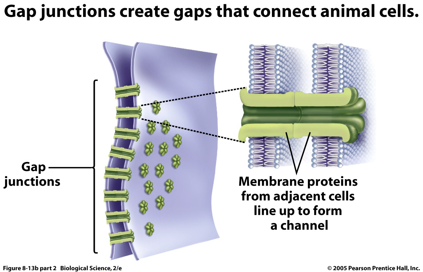
 

2)      Cell membrane

All biological membranes have a common general structure thin film of lipid and protein molecules, held together mainly by non-covalent interactions. Membranes system specificity governed mostly by proteins associated with them.

Molecular properties of the membrane regulate membrane fluidity. Collision interactions are essential for transfer of substrate molecules. In addition, biological membranes are responsible for the transferring of signals; Electron Transport Chain and assembly of multipurpose complexes (transient or longer).

Cell membrane functions include;

  • Permeability barrier
  • Sensory organelle
  • Medium of transport


3)      Cytoplasm

The cytoplasm is an aqueous environment in the cell which aids in the movement of vesicles and organelles, as well as providing an aqueous environment for chemical reactions to take place.


4)      Mitochondria

The structure of the mitochondria is demarked by the description below.

2 membranes

  • fluid-filled space between 2 membranes
  • internal fluid-filled space

Outer Membrane – has large aqueous channels made of a protein called porin – permeable to most small molecules”

Inner Membrane – impermeable to ions and small molecules except by specific membrane transport proteins

Note that the presence of the double membrane feature is to;

 I.  maximize SA on which chemical reactions occur, the inner membrane is folded, forming pockets called cristae.

II. while the inner membrane surrounds the matrix compartment, mitochondrial DNA, ribosomes, enzymes used in ATP production.

 III. and the inter-membrane space between the inner and outer membranes maintains the hydrogen ion diffusion gradients required to generate ATP.

The function of mitochondria is;

  • cellular respiration generate ATP

The greater the demand for energy in a cell, due to the function it is involved in, the more amount of mitochondria present in the cell.  


5)      Golgi Apparatus

The structure of this organelle is;

  • Stack of membranous saccules
  • Serves in the processing, packaging and distributing of molecules

The Golgi apparatus is integral in modifying, sorting, and packaging these macromolecules for cell secretion (exocytosis) or use within the cell. Its functions include;

•modifies proteins delivered from the rough endoplasmic reticulum

•involved in the transport of lipids around the cell

•the creation of lysosomes

•site of carbohydrate synthesis


6)      Endoplasmic Reticulum

This is a membranous network of cisterns (sac-like structures) held together by the cytoskeleton. There are 2 types of ER;

  • Rough ER is studded with ribosomes. The rough endoplasmic reticulum is studded with ribosomes on the cytosolic face. These are the sites of protein synthesis.
  • Smooth ER lacks ribosomes. The smooth endoplasmic reticulum is a smooth network without the ribosomes. The smooth endoplasmic reticulum is concerned with lipid metabolism, carbohydrate metabolism, and detoxification.


7)     Lysosomes

Lysosomes are cellular organelles (vesicles) that contain acid hydrolase enzymes that break down waste materials and cellular debris. They can be described as the stomach of the cell.

8)     Peroxisomes

These are organelles (vesicles) found in all eukaryotic cells. They are involved in the catabolism of very long chain fatty acids, branched chain fatty acids, D-amino acids, polyamines, and biosynthesis of phospholipids. 

Post 24

Post 24… I’ve search and deliberated quite a lot for this one

Biochemistry, sometimes called biological chemistry, is the study of chemical processes within, and relating to, living organisms. By controlling information flow through biochemical signaling and the flow of chemical energy through metabolism, biochemical processes give rise to the complexity of life. Over the last 40 years biochemistry has become so successful at explaining living processes that now almost all areas of the life sciences from botany to medicine are engaged in biochemical research. Today the main focus of pure biochemistry is in understanding how biological molecules give rise to the processes that occur within living cells, which in turn relates greatly to the study and understanding of whole organisms.

Biochemistry is closely related to molecular biology, the study of the molecular mechanisms by which genetic information encoded in DNA is able to result in the processes of life. Depending on the exact definition of the terms used, molecular biology can be thought of as a branch of biochemistry, or biochemistry as a tool with which to investigate and study molecular biology.

Much of biochemistry deals with the structures, functions and interactions of biological macromolecules, such as proteins, nucleic acids, carbohydrates and lipids, which provide the structure of cells and perform many of the functions associated with life. The chemistry of the cell also depends on the reactions of smaller molecules and ions. These can be inorganic, for example water and metal ions, or organic, for example the amino acids which are used to synthesise proteins. The mechanisms by which cells harness energy from their environment via chemical reactions are known as metabolism.

What we take away from this course?

Nothing is as simple as it seems…!!! The simplest process which we usually overlook is actually more complex in its functionality, that the physical activities in which we willing part take in. We a complex individual are made up of numerous simple chemical reactions and relationships which dictate out being. Also, every single process and relationship is interlinked via some means in order to produce working system. 

Image

Nucleotides and Nucleic Acids II

Stability of Nucleic Acid

1)      Hydrogen bonding

Does not normally contribute the stability of nucleic acids

Contributes to DNA double helix, RNA secondary structure

2) Stacking interaction/hydrophobic interaction between aromatic base pairs/bases contribute to the stability of nucleic acids.

It is energetically favourable for the hydrophobic bases to exclude waters and stack on top of each other

This stacking is maximized in double-stranded DNA


Effect of Acid

  • Strong acid + high temperature: completely hydrolyzed to bases, riboses/deoxyrobose, and phosphate
  • pH 3-4 :  apurinic nucleic acids [glycosylic bonds attaching purine (A and G) bases to the ribose ring are broken ], can be generated by formic acid

Effect of Alkali

  • High pH (> 7-8) has  subtle (small) effects  on DNA structure
  • High pH changes the tautomeric state of the bases


Denaturation

Urea (H2NCONH2): denaturing PAGE

Formamide (HCONH2): Northern blot


Step 1: Disrupting the hydrogen bonding of the bulk water solution

Step 2: Hydrophobic effect (aromatic bases) is reduced 

Step 3: Denaturation of strands in double helical structure


C3 Spectroscopic and Thermal Properties of Nucleic Acids

1. UV absorption:

2. Hypochromicity: caused by the fixing of the bases in a hydrophobic environment by stacking, which makes these bases less accessible to UV absorption.

3. Quantitation of nucleic acids

4. Purity of DNA

5. Thermal denaturation/melting: heating leads to the destruction of double-stranded hydrogen-bonded regions of DNA and RNA.

6. Renaturation


DNA Supercoiling

  • Closed circular molecule
  • Supercoiling & energy
  • Topoisomer & topoisomerase

Nucleotides and Nucleic Acids

Nucleotides and Nucleic Acids


Transcription:

Transcription is the first step of gene expression, in which a particular segment of DNA is copied into RNA by the enzyme, RNA polymerase. Both RNA and DNA are nucleic acids, which use base pairs of nucleotides as a complementary language that can be converted back and forth from DNA to RNA by the action of the correct enzymes. During transcription, a DNA sequence is read by an RNA polymerase, which produces a complementary, antiparallel RNA strand. As opposed to DNA replication, transcription results in an RNA complement that includes uracil (U) in all instances where thymine (T) would have occurred in a DNA complement. Also unlike DNA replication where DNA is synthesised, transcription does not involve an RNA primer to initiate RNA synthesis.


 Image

Translation:

In molecular biology and genetics, translation is the process through which cellular ribosomes manufacture proteins. It is part of the process of gene expression. In translation, messenger RNA (mRNA) produced by transcription is decoded by the ribosome to produce a specific amino acid chain, or polypeptide, that will later fold into an active protein. In bacteria, translation occurs in the cell’s cytoplasm, where the large and small subunits of the ribosome are located, and bind to the mRNA. In eukaryotes, translation occurs across the membrane of the endoplasmic reticulum in a process called vectorial synthesis. The ribosome facilitates decoding by inducing the binding of tRNAs with complementary anticodon sequences to that of the mRNA. The tRNAs carry specific amino acids that are chained together into a polypeptide as the mRNA passes through and is “read” by the ribosome in a fashion reminiscent to that of a stock ticker and ticker tape.


 Image

Nucleotide

Nucleotides are biological molecules that form the building blocks of nucleic acids (DNA and RNA) and serve to carry packets of energy within the cell (ATP). In the form of the nucleoside triphosphates (ATP, GTP, CTP and UTP), nucleotides play central roles in metabolism. In addition, nucleotides participate in cell signaling (cGMP and cAMP), and are incorporated into important cofactors of enzymatic reactions (e.g. coenzyme A, FAD, FMN, NAD, and NADP+).


 Image

DNA Replication Base Pairing

Base pairs are the building blocks of the DNA double helix, and contribute to the folded structure of both DNA and RNA. Dictated by specific hydrogen bonding patterns, Watson-Crick base pairs (guanine-cytosine and adenine-thymine) allow the DNA helix to maintain a regular helical structure that is independent of its nucleotide sequence. The complementary nature of this based-paired structure provides a backup copy of all genetic information encoded within double-stranded DNA. The regular structure and data redundancy provided by the DNA double helix make DNA well suited to the storage of genetic information, while base-pairing between DNA and incoming nucleotides provide the mechanism through which DNA polymerase replicates DNA, and RNA polymerase transcribes DNA into RNA. Many DNA-binding proteins can recognize specific base pairing patterns that identify particular regulatory regions of genes.

Intramolecular base pairs can occur within single-stranded nucleic acids. This is particularly important in RNA molecules (e.g. transfer RNA), where Watson-Crick base pairs (G-C and A-U) permit the formation of short double-stranded helices, and a wide variety of non-Watson-Crick interactions (e.g. G-U or A-A) allow RNAs to fold into a vast range of specific three-dimensional structures. In addition, base-paring between transfer RNA (tRNA) and messenger RNA (mRNA) forms the basis for the molecular recognition events that result in the nucleotide sequence of mRNA becoming translated into the amino acid sequence of proteins.


Role:

  • Building blocks of nucleic acids (RNA, DNA)
  • Analogous to amino acid role in proteins


  • Energy currency in cellular metabolism (ATP: adenosine triphosphate)


  • Allosteric effectors


  • Structural components of many enzyme cofactors (NAD: nicotinamide adenine dinucleotide)


  • DNA contains genes, the information needed to synthesize functional proteins and RNAs


  • DNA contains segments that play a role in regulation of gene expression (promoters)


  • Ribosomal RNAs (rRNAs) are components of ribosomes, playing a role in protein synthesis


  • Messenger RNAs (mRNAs) carry genetic information from a gene to the ribosome


  • Transfer RNAs (tRNAs) translate information in mRNA into an amino acid sequence


  • RNAs have other functions, and can in some cases perform catalysis


Nucleosides

Nucleosides are glycosylamines consisting of a nucleobase (often referred to as simply base) bound to a ribose or deoxyribose sugar via a beta-glycosidic linkage. Examples of nucleosides include cytidine, uridine, adenosine, guanosine, thymidine and inosine.


Nucleic Acids

Nucleic acids are large biological molecules essential for all known forms of life. They include DNA (deoxyribonucleic acid) and RNA (ribonucleic acid). Together with proteins, nucleic acids are the most important biological macromolecules; each is found in abundance in all living things, where they function in encoding, transmitting and expressing genetic information.


RNA

  • rRNA

RNA + protein form ribosomes


  • mRNA

carries DNA code to cytosol for protein synthesis on the ribosomes

read in triplets called codons (5’ to 3’)


  • tRNA

carries amino acids to mRNA and matches up by base-pairing of triplet anti-codon with mRNA codon triplet

Another round of MCQ’s

1) At the end of glycolysis, how much ATP is generated?

(A) 1
(B) 2
(C) 3
(D) 4
(E) 5


2) The smallest free-living cell is a bacteria called:

(A)  Proteobacteria

(B)  Verrucmicobia

(C)  Dialister Bacteria

(D)  Actinbacteria

(E)  Cyanobacteria

Enzyme 4

The Lineweaver–Burk plot (or double reciprocal plot) is a graphical representation of the Lineweaver–Burk equation of enzyme kinetics, described by Hans Lineweaver and Dean Burk in 1934.

Image 


The Michaelis–Menten kinetics is one of the simplest and best-known models of enzyme kinetics. It is named after German biochemist Leonor Michaelis and Canadian physician Maud Menten. The model takes the form of an equation describing the rate of enzymatic reactions, by relating reaction rate  to , the concentration of a substrate S.

 Image

The Lineweaver–Burk plot was widely used to determine important terms in enzyme kinetics, such as Km and Vmax, before the wide availability of powerful computers and non-linear regression software. The y-intercept of such a graph is equivalent to the inverse of Vmax; the x-intercept of the graph represents −1/Km. It also gives a quick, visual impression of the different forms of enzyme inhibition.

The double reciprocal plot distorts the error structure of the data, and it is therefore unreliable for the determination of enzyme kinetic parameters. Although it is still used for representation of kinetic data, non-linear regression or alternative linear forms of the Michaelis–Menten equation such as the Hanes-Woolf plot or Eadie–Hofstee plot are generally used for the calculation of parameters.

When used for determining the type of enzyme inhibition, the Lineweaver–Burk plot can distinguish competitive, non-competitive and uncompetitive inhibitors. Competitive inhibitors have the same y-intercept as uninhibited enzyme (since Vmax is unaffected by competitive inhibitors the inverse of Vmax also doesn’t change) but there are different slopes and x-intercepts between the two data sets. Non-competitive inhibition produces plots with the same x-intercept as uninhibited enzyme (Km is unaffected) but different slopes and y-intercepts. Uncompetitive inhibition causes different intercepts on both the y- and x-axes but the same slope.